Premium Masterclass
Full HD Masterclass AccessThis full 4k surgical video is locked. Join the Surgeon Pro plan to watch this procedure and the entire video library.
Unlock Full Access
Already a member? Log in
Near Live Surgery
14m 00s
Full Masterclass
Hysteroscopic Adenomyoma Resection (NL0067)
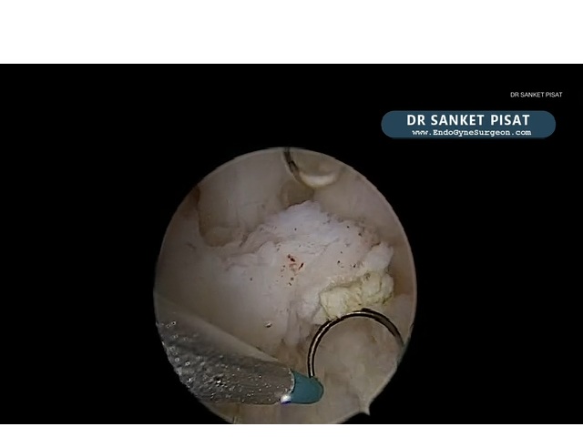
This video demonstrates hysteroscopic resection of a small, predominantly intramural focal adenomyoma in a patient with infertility and two previous failed IVF attempts.
Initial diagnostic hysteroscopy reveals only subtle abnormalities, including focal endometrial irregularity, increased vascularity and a minimal cavity bulge corresponding to the ultrasound findings.
The surgeon reduces intrauterine distension pressure to improve localization of the lesion, which does not protrude significantly because of its intramural position.
Hysteroscopic scissors are used to circumferentially incise and progressively separate the adenomyoma from the surrounding myometrium, encouraging it to project into the cavity.
An 18 Fr bipolar resectoscope is subsequently introduced to remove the mobilized lesion using controlled, gentle cutting movements.
The surgeon differentiates residual adenomyotic tissue from normal myometrium by their appearance, emphasizing the importance of avoiding excessive deep resection and uterine perforation.
The procedure concludes with near-complete macroscopic removal of the lesion, specimen retrieval and histopathological confirmation of adenomyoma, allowing the patient to proceed with further fertility treatment.
Instructor

Dr. Sanket Pisat
Lead InstructorPioneer in Gynaecological Endoscopy, bringing 15+ years of surgical excellence.
View Profile